In spite of the heavy rain, many alumni joined us for an alumni donor appreciation crawfish boil on the afternoon of
A Nature Communications Paper by Physicist Michalis Charilaou
Sun, 07/09/2023 - 7:04pm
Top Stories
Students Sink Teeth into New Science Museum Exhibit
A new exhibit opening 1 May 2026 at the University of Louisiana at Lafayette Science Museum is putting student creat
An interesting paper by Michalis Charilaou of our Department of Physics, along with six of his colleagues has recently appeared in the prestigious journal, Nature Communications. You may read the paper at the following link Direct observation of tensile-strain-induced nanoscale magnetic hardening.
The paper is a contribution to the long quest to clarify the link between structure and magnetism in matter. Research into this link has a long history, dating back to the 19th century when J. Joule and E. Villari discovered magnetostriction and magnetoelasticity. Since then, stress and stress annealing have found applications in the sensing, control and enhancement of magnetic properties in a wide range of materials and devices, ranging from magnetic random-access memory to energy harvesting and biomedicine. Despite the long history, the physics of magnetoelasticity on the nanoscale remains poorly understood.
In this study, the authors performed cutting-edge, high-resolution imaging experiments by means of electron microscopy and holography, as well as micromagnetic simulations and they discovered that when stress is applied on a thin ferromagnetic plate of Nickel, the magnetic domains in the plate change shape, as seen in the figure here, which shows the magnetic field inside the sample. By comparing these experiments with simulations, they found that this changing texture of the magnetization in the material is the result of strain-induced magnetic anisotropy, i.e., a preferential direction of the magnetization is induced in the sample because of strain. The mechanism of this induced anisotropy lies in the small change of the distances between the atoms when the material experiences strain, where a small change in the atomic scale can have a dramatic scale in the nanoscale. Importantly, this change in the magnetic state has implications on the macroscopic properties of the material as well; its response to an external field will change. This suggests that thin ferromagnetic plates can be used as sensors for mechanical strain via magnetic signals.
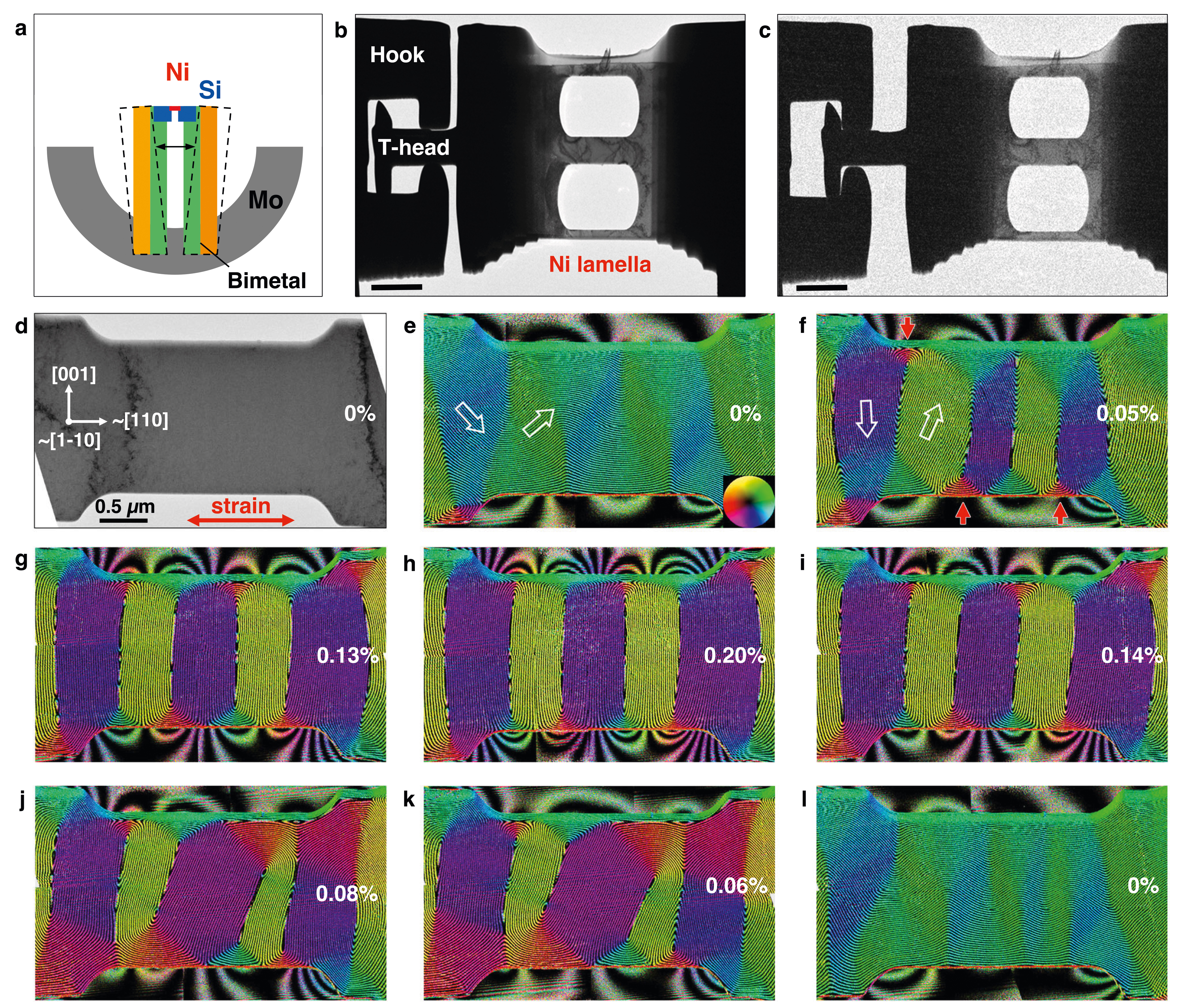
Image caption. Direct measurement of magnetostriction in a Ni nanostructure.
- Figure a: Schematic diagram of the bimetallic deformation device at room temperature and elevated temperature (dashed lines).
- Figures b and c: BF TEM images of the device in b unstrained and c strained conditions. The scale bar is 2 μm. In order to reduce the temperature effect, the distance between the hooks and the T-head was ∼1 μm, enabling the straining experiment to be carried out below 60 °C.
- Figure d: BF TEMimage of a single crystalline Ni nanostructure recorded before a second tensile cycle. The white arrows mark the crystallographic orientation of the sample. Strain was applied in the horizontal direction (marked by a double-headed red arrow) at the levels indicated in each subsequent image. The error is estimated to be ±0.04%.
- Figures e - l: Real space magnetic induction maps recorded during a straining and release cycle using off-axis EH. The direction of the projected in-plane magnetic induction is visualized both according to the color wheel shown in (e) and using hollow white arrows. The contour spacing is 2π/3 radians. The small red arrows in (f) mark the positions of magnetic flux closure domains.